Graduate Microanatomy, 1998
Graduate Microanatomy, 1998
|
Date page was last edited 06/06/04 |
Lab Exercises: EAR The lab exercises will focus on structures in the inner ear, particularly those involved with the sensory cells. There are several excellent overview diagrams in Bloom and Fawcett. Consult page 92, Figure 35-1 for an overview of the Ear canal and middle ear. 1. Trace the sound waves down the ear canal into the tympanic cavity. What is the function of the auditory ossicles, tympanic membrane, tensor tympani, and stapedius muscles?
2. How are sound waves transformed into pressure on the fluid of the inner ear?
On slide 7, find the bony labyrinth. Most of the slide shows the snail like COCHLEA. Look at the cochlea with the lowest power. Consult figure 35-9 for a vew of the cochlea. The coiled structure is also appreciated by the drawing in Figure 35-7 where the organ of Corti is seen diagrammatically in black along the wall of the cochlea. This photograph shows an overview of the cochlea. 3. What fluid flows through the membranous labyrinth? What fluid flows through the bony labyrinth?
The membranous labyrinth is divided into three chambers in a given section through one of the cochlear turns. These can be identified with the help from Figure 35-10. The following photograph is from your class slide 7. 4. Identify each of the chambers and the fluid in each chamber.
Look at the contents of the Scala media. The ORGAN OF CORTI sits on the BASILAR MEMBRANE. Find one in your class slide set that has a fairly intact neuroepithelium. Use Figures 35-12 and 35-13 to help you find some of the structures in the organ of Corti.
4. Identify as many parts of the organ of Corti in the above photo as you can.
5. How do the hair cells get stimulated by the sound waves? What specializations are on their surface and what is the role of the tectorial membrane?
6. Look at the difference in supportive cells adjacent to the inner and outer hair cells. How could this arrangement aid in the contactile ability of the outer hair cells?
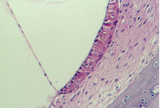
Along the wall of the cochlea, there is a band of stratified epithelium called the stria vascularis. It contains a plexus of capillaries which even extend up into the epithelium. The following photograph shows a high magnification of the stria vascularis. You can see Reissner's membrane at one end, dividing the scala media from the scala vestibuli.
7. What is the function of the stria vascularis?
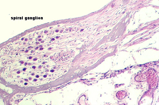
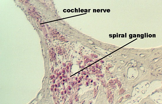
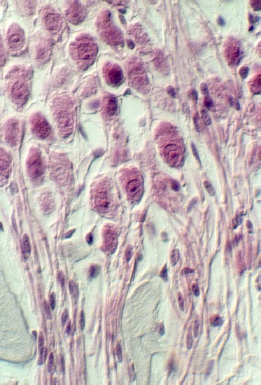
The following photograph of the organ of Corti show the cochlear nerve passing from the hair cells through the bone. It passes to the SPIRAL GANGLION CELLS which are seen well in most preparations.
The spiral ganglion is seen in the following photographs.
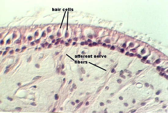
Review figure 35-3 to gain an appreciation of the membranous labyrinth for this region of the inner ear. Your slides 7 or 99 may have examples of neuroepithelial sensory structures in one or more of these regions. They may have been distorted, so you will have to rely on lecture notes and your text to orient yourself. This photograph shows a view of the crista ampullaris. Use Figure 35-3 to see a drawing of this structure. Higher magnifications are shown in the next photographs.
. Note: Even though the above figure is labeled afferent nerve fibers, one cannot distinguish these at the light microscopic level. Find a crista ampullaris in your ear slides. Notice the position of the VESTIBULAR GANGLION (SCARPA's GANGLION) and its relationship with the cochlear division of Cranial Nerve VIII. 8. What are the distinguishing features of a Type I and Type II hair cell at the electron microscopic level? Which hair cells can be identified in these photos and why?
You may also be able to see an example of a macula from either the saccule or utricle in one of your ear slides. Use Figure 35-8 in your text as a guide. The following photographs show different magnifications of a macula. You may be able to distinguish the SACCULE which is embedded in the bone, just adjacent to the cochlea. The relationship of the utricle within the inner ear has been distorted. Identify HAIR CELLS and what is left of the OTOLITH MEMBRANE.
Return to top of page
| Course Design | Learning Aids | Learning modalities | |
 Slides 7 and 99 show views of the inner ear which may show one or more of the
sensory cell complexes. In order to understand how the inner ear is organized, compare
Figures 35-1 and 35-3 in Bloom and Fawcett. Figure 35-1 shows how the membranous
parts of the inner ear are embedded in the BONY LABYRINTH and figure 35-3 is a drawing of
the MEMBRANOUS LABYRINTH.
Slides 7 and 99 show views of the inner ear which may show one or more of the
sensory cell complexes. In order to understand how the inner ear is organized, compare
Figures 35-1 and 35-3 in Bloom and Fawcett. Figure 35-1 shows how the membranous
parts of the inner ear are embedded in the BONY LABYRINTH and figure 35-3 is a drawing of
the MEMBRANOUS LABYRINTH. 


 Semicircular Ducts; Utricle and Saccule