Graduate Microanatomy, 1998
Graduate Microanatomy, 1998
|
Date page was last edited 06/06/04 |
Lab exercises:
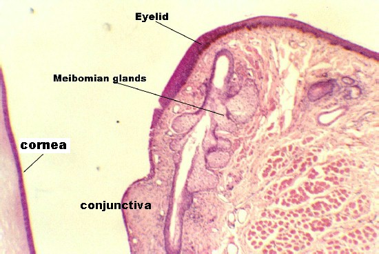
Eyelid and Lacrimal Gland First, orient yourself to slide 97. Hold slide 97 up to the light and, using the diagram in Figure 34-2, locate the front and back of the eye. Find the lens. That chamber in front of the lens is the ANTERIOR CHAMBER. The chamber beside and behind the lens is the POSTERIOR CHAMBER. It is filled with the VITREOUS BODY. The anterior border of the anterior chamber is the CORNEA. Once you have identified this region, then find the EYELIDS which are anterior to the cornea. With the microscope, one can see that the skin of the outer part of the eyelid is lined by cornified stratified squamous epidermis. There are hair follicles which house the eyelashes. The sebaceous glands that secrete into the eyelashes are the MEIBOMIAN GLANDS (tarsal glands). Find these regions with the following photograph as a guide. The photo below also shows the cornea to the left of the eyelid. The region under the eyelid is called the CONJUNCTIVA. Inside the eyelid can be seen bundles of the ORBICULAR MUSCLE
The figure below shows a higher magnification of Meibomian glands emptying into the hair follicle of an eyelash.
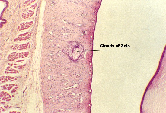
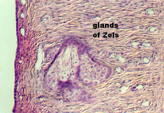
Small sebaceous and sweat glands can also be seen in the dermis. The sebaceous glands that do not empty into hair follicles are called the GLANDS OF ZEIS. This is shown in the following two photographs. Note that it is emptying into the space between the conjunctiva (in which it sits) and the cornea (shown to the right
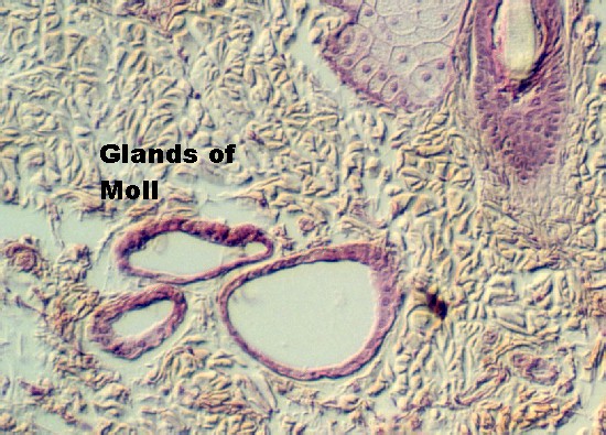
The small sweat glands in the dermis can be hard to find. They also have a special name. They are called the GLANDS OF MOLL. The following photograph illustrates an example near some MEIBOMIAN GLANDS.
The orbicular muscle is illustrated in the next photograph. It is striated muscle.
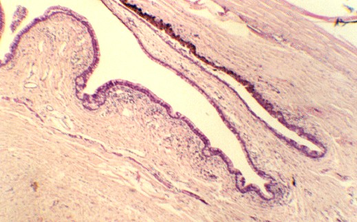
The conjunctiva continues as the anterior covering of the CORNEA. Move to the edge of one of the eyelids and examine the junction at a region called the CONJUNCTIVAL FORNIX. This junction is shown below. The cornea is running along top, upper right hand cornea. The conjunctiva with its Goblet cells is facing the cornea and distinguished by multiple folds.
The following figure shows the conjunctiva with its Goblet cells facing the pigmented epithelium of the cornea seen near the junction.
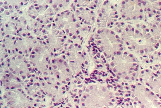
Lacrimal Gland Another source of lubrication for the eye comes from the lacrimal gland (tear gland). Look at slide 98. Two views are in your slide sets. One contains a large portion of the gland and the other contains only a small portion. The lacrimal gland is a serous goand. It is organized into alveoli, like the parotid gland. Adipose tissue may be mixed with the glandular cells. The following illustrations show the gland cells.
| Course Design | Learning Aids | Learning modalities | |