

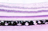

Hold your slide of the eye up to the light and identify the different regions.Find the lens. That chamber in front of the lens is the ANTERIOR CHAMBER. The chamber beside and behind the lens is the POSTERIOR CHAMBER. It is filled with the VITREOUS BODY. The anterior border of the anterior chamber is the CORNEA. Once you have identified this region, then find the EYELIDS which are anterior to the cornea.
Eyelids

With the microscope, one can see that the skin of the outer part of the eyelid is lined by cornified stratified squamous epidermis. There are hair follicles which house the eyelashes. The sebaceous glands that secrete into the eyelashes are the MEIBOMIAN GLANDS (tarsal glands). Find these regions with the photos as a guide. The above photo also shows the cornea to the left of the eyelid. The region under the eyelid is called the CONJUNCTIVA. Inside the eyelid can be seen bundles of the ORBICULAR MUSCLE
The figure below shows a higher magnification of Meibomian glands emptying into the hair follicle of an eyelash.

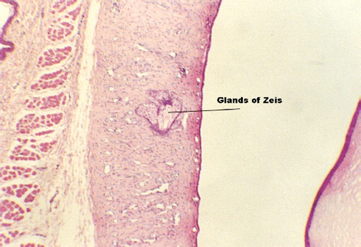
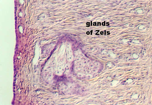
Small sebaceous and sweat glands can also be seen in the dermis. The sebaceous glands that do not empty into hair follicles are called the GLANDS OF ZEIS. This is shown in the following two photographs. Note that it is emptying into the space between the conjunctiva (in which it sits) and the cornea (shown to the right
The small sweat glands in the dermis can be hard to find.
They also have a special name. They are called the GLANDS OF MOLL. The
following photograph illustrates an example near some MEIBOMIAN GLANDS.

The orbicular muscle is illustrated in the next photograph. It is striated muscle.
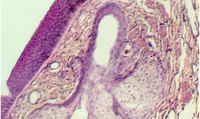
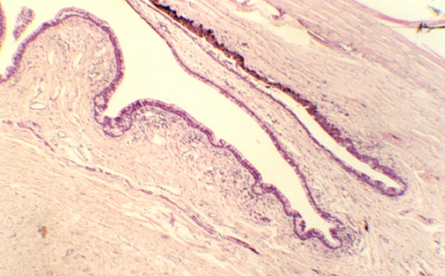
The conjunctiva lines the inside of the eyelids, extending to cover the sclera (bottom left epithelium in following figure). Its epithelium varies from non-keratinized stratified squamous epithelium to stratified cuboidal or columnar epithelium, with Goblet cells. Move to the edge of one of the eyelids and examine the junction at a region
called the CONJUNCTIVAL FORNIX. This junction is shown below. The cornea is
running along top, upper right hand cornea. The conjunctiva is
facing the cornea and distinguished by multiple folds.
\
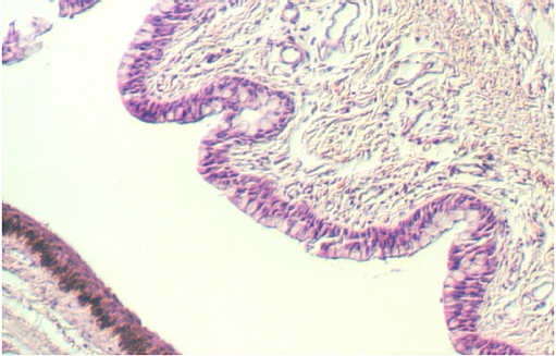
The following figure shows the conjunctiva as a stratified columnar or cuboidal epithelium (top, right) containing numerous Goblet cells.
Lacrimal Gland
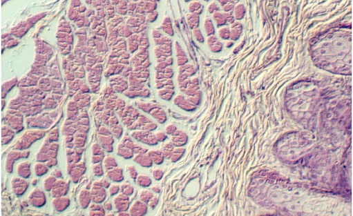
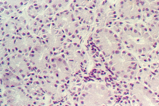
Another source of lubrication for the eye comes from the lacrimal gland (tear gland). The lacrimal gland is a serous gland. It is organized into alveoli, like the parotid gland. Adipose tissue may be mixed with the glandular cells. The following illustrations show the gland cells.

http://microanatomy.net/Eye/eyelids_and_lacrimal_glands.htm
Gwen V. Childs, Ph.D., FAAA
Department of Neurobiology and Developmental Sciences
University of Arkansas for Medical Sciences
4301 W. Markham, Slot 510, Little Rock, AR 72205
For questions or concerns, send email to this address
