Graduate Microanatomy, 1998
Graduate Microanatomy, 1998
|
Date page was last edited 06/06/04 |
Lab exercises:
Eye An overview of the cornea proper is shown to the left. The thicker epithelium is facing anteriorly (left). The outside of the eye would be to the left. Then, one can see the cornea proper. Finally Descemet's membrane and the mesothelium facing the anterior chamber is evident. The following photograph shows these structures in greater detail. Compare it with Figure 34-3 from your text. Bowman's membrane is actually the first part of the Cornea proper and sits just under the epithelium which is facing outward. Descemet's membrane is just inside the mesothelium, which faces the anterior chamber.
Move your slide into the anterior chamber. The first structures you may encounter on each side of the LENS constitutes the IRIS. This is heavily pigmented along the surface facing the lens. Not all sections go through the pupil. The following photographs show the iris and its relationship to the lens. In the photographs below, the center of the eye of the pupil is to the right. Inside the iris can be seen bundles of muscle. 1. What type of muscle is this and what is its function? How is it innervated?
LENS The lens may be torn in your slides. However, one can see lens epithelium immedicately under the capsule. Also, if you focus up and down, you can see the equatorial arrangement of lens fiber nuclei. At any rate, the fiber organization can be seen somewhat. The following photograph shows the lens epithelium, the fibers, and the nuclei.
2. What structures in the eye control the lens?
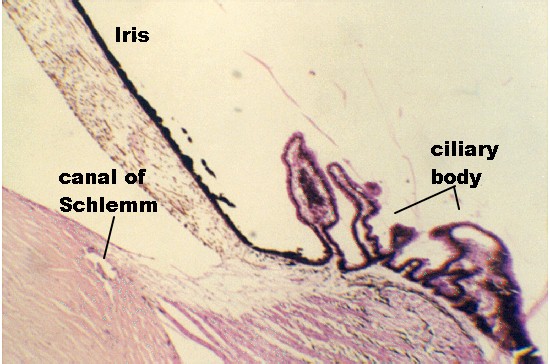
CILIARY BODY Move to the iridocorneal angle or junction and the CILIARY BODY can be seen. It has several processes with pigmented epithelium. Projecting from these processes are fibers called "zonule fibers" that connect with the lens. The following photographs illustrate the CILIARY PROCESSES and Zonule fibers.
This region also separates the anterior and posterior chambers of the eye. At the iridocorneal junction, one can see the canal of Schlemm, which almost looks like a tear in the corneal connective tissue. 3) In the following photograph, label the anterior chamber and posterior chambers using Figure 34-9 as a guide.
4) What is the function of the Canal of Schlemm?
RETINA and the VASCULAR TUNIC Begin along the inside of the ciliary body and follow the pigmented epithelial cells along the inside of the wall of the eye. This inner layer is called the PARS CAECA of the retina. The retina begins as the PARS OPTICA at a region called the ORA SERRATA. Find this region in your section.
In this region, you can also see the pigmented choroid coat underneath the lining epithelium. It contains three layers: the outer, vessel layer, the middle, capillary layer (choriocapillary layer), and Bruch's membrane, lying just under the pigmented epithelium. The vascular layer with its large vessels can be appreciated in the photograph below, showing an overview of the retina. 5) What components make up Bruch's membrane?
The lowest layer shown in the photo above is the sclera. This is the connective tissue sheath that supports the eyeball. It also contains elastic fibers and pigmented cells Chapter 35, Bloom and Fawcett 1. Briefly, define the following structures: Auricle, external acoustic meatus, middle ear, auditory ossicles. 2. What is the function of the auditory ossicles and the tympanic membrane? How are the sound waves transformed into pressure waves. 3. In the internal (inner) ear, what is the difference between the bony labyrinth and the membranous labyrinth? Where is endolymph and perilymph and what cells produce these fluids? 4. What is the function of the semicircular ducts (canals)? What sensory cells perform this function and where are they located in the canals? 5. Describe the cristae ampullaris and its function. How are Hair cells designed to support their function? 6. Define the function of the utricle and saccule and its sensory cells. Where are they located? 7. Be able to draw or identify the following parts of the cochlea: modiolus, basilar membrane, vestibular membrane, scala vestibuli, scala tympani, scala media, stria vascularis. 8. Draw a cross section through the scala media and label: vestibular membrane, endolymph, basilar membrane, stria vascularis, Organ of Corti, tectorial membrane. 9. In the Organ of Corti, define the structure and function of: the different types of supportive cells; the sensory cells. Where do the nerve fibers collect before they travel to the brain. What is the role of the endolymph and tectorial membrane? 10. Trace the cochlear nerve fibers to their origin....where are the cell bodies? Distinguish Scarpa's ganglion and the Spiral ganglion? Name the nerve that is formed from fibers from these cell bodies.
| Course Design | Learning Aids | Learning modalities | |