

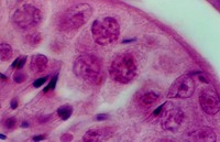


The Prostate epithelium looks cuboidal or pseudostratified low columnar in spots. Inside the glandular lumena you may see laminated bodies known as concretions. These are evident in the following photograph and help identify the organ as the prostate.
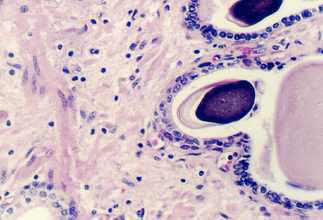
In the connective tissue there are bundles of smooth muscle which stand out
because of their homogeneous cytoplasm. Find smooth muscle in the above photograph
or the one below. What components of semen are contributed by the prostate?
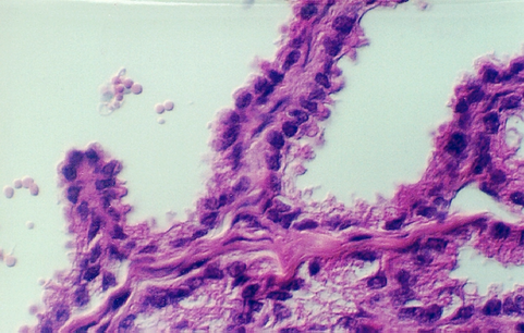
The lumen of this gland is thrown into many folds that appear to branch. This creates a labyrinth of channels, tubes, and pockets into which the secretory product flows. The epithelium shown in the photos below may be simple columnar with patches of pseudostratified epithelium. The appearance of the epithelium is variable and dependent on age and the level of androgen production. One can see some lipochrome pigment in the epithelium, which is present in elderly males. Look at the wall of the gland and again, there are numerous bundles of smooth muscle.


What does the seminal vesicle contribute to the semen?

http://microanatomy.net/Male_Reproductive/prostate_and_seminal_vesicle.htm
Gwen V. Childs, Ph.D., FAAA
Department of Neurobiology and Developmental Sciences
University of Arkansas for Medical Sciences
4301 W. Markham, Slot 510, Little Rock, AR 72205
For questions or concerns, send email to this address
