Graduate Microanatomy, 1998
Graduate Microanatomy, 1998
|
Bone and cartilage study guide Lab Exercises: Bone Development: Endochondral Bone Development: Intramembranous
Date page was last edited 06/06/04 |
Laboratory Exercises: Cartilage
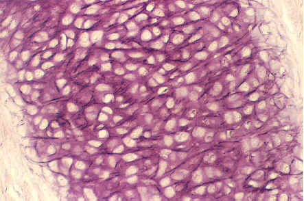
Hyaline Cartilage: Trachea In slide 45, Find the C-Shaped ring in the connective tissue of trachea. This is a good example of hyaline cartilage. Look at the cell layers surrounding the cartilage. The outermost layer is the perichondrium. Moving inside the hyaline cartilage, note the chondrocytes embedded in their lacunae in the homogeneous matrix.
Hyaline Cartilage: Trachea This is a higher magnification of hyaline cartilage in the trachea showing the chondroblasts in the perichondrium and the chondrocytes in the lacunae and the matrix. Some cells may appear to be in clusters in an individual lacuna. These are called "isogenous groups". Higher magnifications are seen below.
Hyaline Cartilage: Trachea
Hyaline Cartilage: Trachea
Elastic Cartilage (ear of newborn) Look at your slide 19. It is stained with a special elastica stain to bring out the elastic fibers. This cartilage differs from hyaline cartilabe because it contains a considerable amount of elastic fibers which are stained bluish black. They traverse the matrix in all directions. The chondrocytes are invisible because no counterstaining was done in this preparation. The lacunae may appear empty. You may also be able to see hairy skin of the ear. A higher magnification is seen in the following photograph. This photo was taken of tissue that was counterstained and you can see the chondrocytes in the matrix. Elastic Cartilage
Fibrocartilage
Fibrocartilage Look at slide 10 in your set. This tissue was taken from an intervertebral disk. There are many artifacts which developed during the processing (tearing, compression, etc) Nevertheless, it is possible to observe several characteristic features of fibrocartilage. The above photograph (not taken of your slide) also shows fibrocartilage. Note the abundance of collagenous fibers and fiber bundles in the ground substances. These are interwoven in a dense feltwork. Scattered throughout the section are chondrocytes which appear very small and sparse compared with elastic and hyaline cartilage. You may see them only by their dense blue nuclei. They are in ill-defined lacunae which may have a yellowish coloration in your slide. In the photo above, you can only see the dense blue-purple spots which are the nuclei.
Exercises: 1. Cartilage has two ways of growing: appositional and interstitial. Briefly describe each type of growth. Also, identify where each type of growth occurs on on one of the above photographs. Appositional:
Interstitial:
2. Why is the chemistry of the cartilage matrix so important to the health and survival of the chondrocyte?
3. Cartilage can be used in transplants without immunosuppressants. What unique property might allow it to be transplanted so easily?
4. Where is fibrocartilage found and what properties allow it to perform its function?
Return to top of page | Course Design | Learning Aids | Learning modalities | |