Graduate Microanatomy, 1998
Graduate Microanatomy, 1998
|
Bone and cartilage study guide Lab Exercises: Bone Development: Endochondral Bone Development: Intramembranous
Date page was last edited 06/06/04 |
Laboratory Exercises:
Endochondral
Endochondral Bone Formation Endochondral bone formation begins from a cartilage model during fetal development. This photograph shows a section through a hand of a fetus. Note the fingers (long bones) and the wrist bones which are the smaller rounder bones. You can begin to see the earliest stages of bone development in the long bones. Can you find where the primary ossification center will be in one of the fingers in the above photograph? It is more obvious in the following photograph which is a sagittal section through a fetal foot. Here, you can see the actual ossification center in the phalangeal bone.
Endochondral Bone Formation Look at slide 20 and find a region showing two adjacent developing bones and a joint cavity. Most of your slides have both primary (diaphyseal) and secondary (epiphyseal) ossification centers. If your slide only shows the secondary center, borrow your neighbor's slide. Note, the joint cavity is lined by hyaline cartilage. There is no perichondrial covering. The joint capsule also is seen. It is composed of dense connective tissue. In the above photo (not from your slide), you can see the hyaline cartilage to the left and the developing bone to the right. In the following slides/exercises, we will identify each zone. In your slide, look for the Epiphyseal plate which is composed of Hyaline cartilage inbetween the primary and secondary ossification centers.
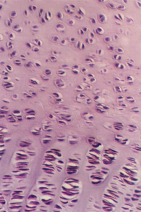
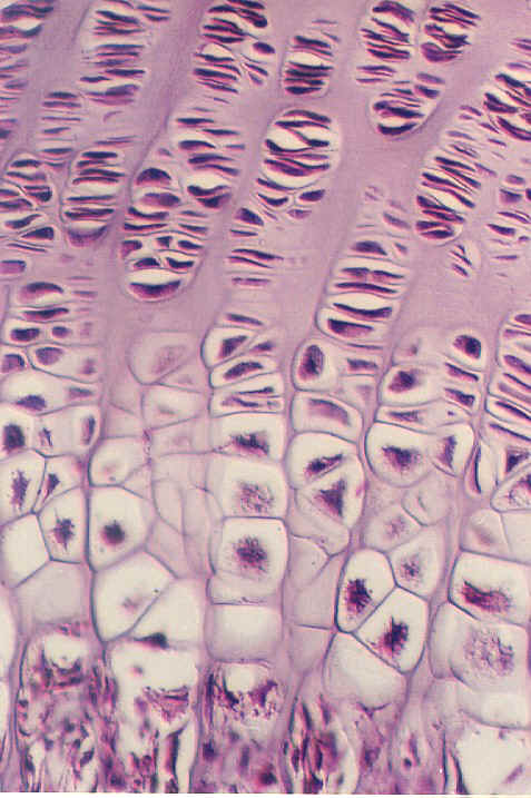
Find the primary center of ossification and then look for the various zones of cartilage in the epiphyseal plate in your slide. Use the photo as a guide. The top zone consists of hyaline cartilage. This is called the Zone of Reserve cartilage (RZ on the photo to the left.) The next zone shows the chondrocytes proliferating. They look like they are stacked up in parallel disks. This is called the Zone of Chondrocyte proliferation. The next zone shows chondrocyte enlargement in their lacunae. This is called the zone of hypertrophy. The cartilage matrix becomes calcified in the zone of calicified cartilage and the chondrocytes die. The only remaining tissue is the calcified cartilage matrix. The osteoblasts then invade the area (with blood vessels) and begin to lay bone on the calcified cartilage matrix. In this photograph, you can see chondrocyte death and the purple matrix (at the bottom) which is beginning to be invated by osteoblasts. This forms the first bone spicules.
This photo shows higher magnifications of which zones of endochondral bone formation? 1. Label each
2. Which zones of endochondral bone formation are shown in this photograph? Label each.
3. What is the major function of the Zone of proliferation?
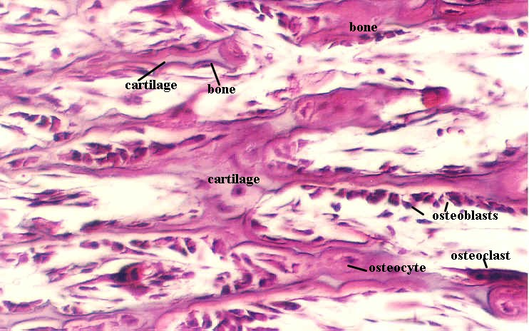
In your slide 20, you may see the secondary ossification center in the epiphysis (end of the long bone). The proliferation zone is not conspicuous, because there is no growth in length. You can see some of the zones of endochondral bone formation, including the hypertrophy. Look also at the periosteum along the diaphysis. Along this area you can see a periosteal collar. This bone strengthens and supports the developing model. However, it is laid down by Intramembranous bone formation. Continue to look at slide 20 with a higher power objective and examine the developing bone spicules. Note areas of purple in the center surrounded by areas of red. 4. What does the purple layer represent?________________________________________ In your slide 20 and the following photographs, you can see the layer of osteoblasts that appear like a layer of cubes on the surface of the spicules. They are producing bone and becoming embedded in it. After they are completely surrounded by bone, they become osteocytes. Find both cell types in the spicules of your bone slide.
Endochondral Bone Formation: Early developing spicules
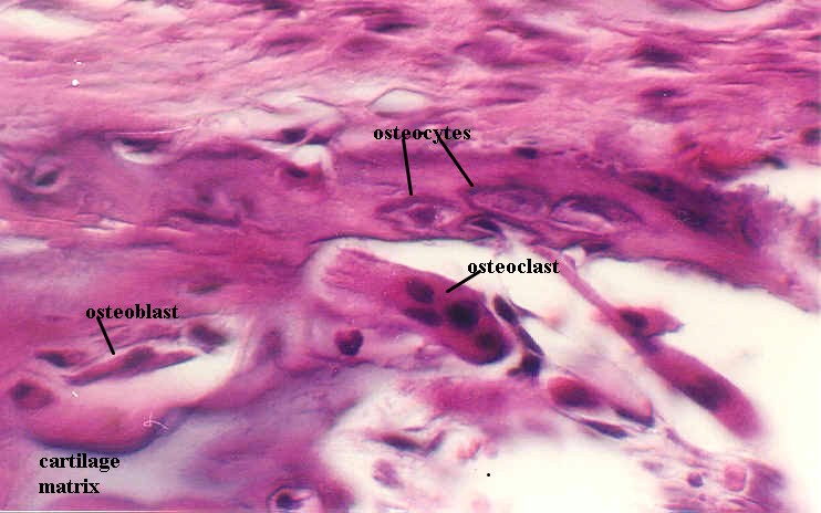
Bone Remodeling As the spicules form, they also become remodeled, a process which continues as long as bone is living. The remodeling starts with osteoclasts, which are multinucleated cells that have a highly eosinophilic cytoplasm. They begin to digest the bone matrix and as they do, they form a depression, called a Howship's lacuna. The osteoblasts then take over and lay down new bone in the direction of the remodeling. Thus, bone can be remodeled to withstand stresses, exercise, etc. In early bone development it is remodeled to create marrow spaces and maintain the proper shape of the bone. Find an osteoclast in your slide 20. Osteoclasts can also be seen in the following photographs. The first of the series shows a somewhat elongated osteoclast (note the 4 nuclei) in Howship's lacuna and cellular processes can be visualized pointing towards the bone. You can also see osteocytes in their lacunae in the bone itself. The second of these photographs shows a rounder osteoclast.
Endochondral bone formation: Remodeling and the osteoclast
Exercises: 5. What is the origin of the osteoclast?
6. What role does periosteum play in bone development, remodeling, and repair?
Return to top of page | Course Design | Learning Aids | Learning modalities | |