 Graduate Microanatomy, 1998
Graduate Microanatomy, 1998
|
Bone and cartilage study guide Lab Exercises: Bone Development: Endochondral Bone Development: Intramembranous
Date page was last edited 06/06/04 |
Laboratory Exercises: Intramembranous Bone Development
Flat bones, like the bones of the skull or jaw bone, another type of bone formation does not begin with a cartilage model. Instead, dense areas of mesenchymal cells transform and begin to lay down bone around themselves, forming early spicules. The above photograph shows trabeculae from fetal skull which have developed via intramembranous bone formation. Look at slide 23, which is a section of developing jaw. The class set contains two different slides. One is a coronal section through the lower half of the head of a 120 mm fetus. You can see the nasal septum and a portion of the devloping maxilla and mandible. You can also see the tongue. The chin is on the right side of the section. In a later lab, you will use this slide to study tooth development. At this point, look for bony spicules, using the photos below as a guide. Note the regions of condensed mesencyme near the spicules. These mesencymal cells have transformed into osteoprogenitor cells and as they produce bone, they become osteoblasts. Osteoprogenitor cells will divide and form new osteoblasts. rows of osteoblasts can be seen on the outer surface of the spicules. Also, you may see numerous osteocytes in the spicule itself.
This higher magnification shows a bony spicule and the row of osteoblasts. You can also identify osteocytes.
Note the fine bone deposition around the cells in the spicule to the right.
Return to top of page | Course Design | Learning Aids | Learning modalities | |
 Intramem-
 This photo shows the earliest development of bone at one end of the
spicule. You can see a fine layer beginning to surround the osteoblast.
This photo shows the earliest development of bone at one end of the
spicule. You can see a fine layer beginning to surround the osteoblast. 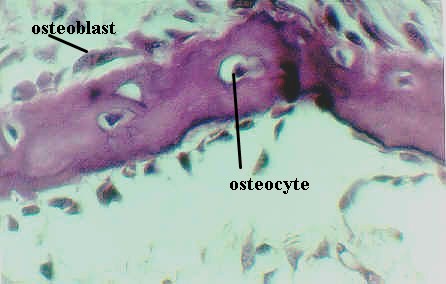
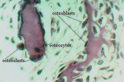 Another higher magnification shows two small osteoclasts on the surface of the
spicule. Thus, bone remodeling is proceeding in intramembranous bone formation as well.
Another higher magnification shows two small osteoclasts on the surface of the
spicule. Thus, bone remodeling is proceeding in intramembranous bone formation as well.