 Graduate Microanatomy, 1998
Graduate Microanatomy, 1998
|
Digestive system study guide Lab Exercises:
Date page was last edited 06/06/04 |
Lab Exercises: Esophagus
Look at slide 50 in your slide set. This is a cross section of the esophagus. Note the NON-KERATINIZED STRATIFIED SQUAMOUS EPITHELIUM lining the lumen. Just under the epithelium, is connective tissue, called the LAMINA PROPRIA. Together, the Epithelium and the lamina propria are called the MUCOSA.
1. Label: epithelium, lamina propria, and muscularis mucosa on this figure
2. Label the SUBMUCOSA
Underneath the submucosa are two muscle layers. The inner layer is a circular layer, cut in cross section in this view. The outer layer is running longitudinally. Together, these muscle layers are called the MUSCULARIS EXTERNA. You can see striated muscle in the muscularis of the upper third of the esophagus. The remainder of the wall consists of smooth muscle. 3. Can you see striated muscle in the figures below, or in your slides? 4. Label the inner and outer muscle layers.
The outermost layer of the esophagus is the ADVENTITIA. It may not be present or abundant in your slide. It consists of loose connective tissue with numerous blood vessels and nerves. In-between the muscle layers, you may see collections of nerve fibers and neuronal cell bodies. This is the AUERBACH's PLEXUS which contains nerves that supply motor control to the muscle and glands in the organ. They are part of the autonomic nervous system. If you don't find any in your esophagus slide, you will no doubt see them in later sections through the GI tract. MEISSNER"s PLEXUS is more difficult to find and recognize. It is dispersed within the connective tissue of the submucosa. Do not spend a lot of time trying to find it.
Look at slide 42, which is through the Gastroesphageal junction. This section is a longitudinal section, so the muscle groups will be running differently. The slide shows the abrupt change in the epithelium from stratified squamous (esophagus) to simple columnar (stomach). It also shows that the muscularis mucosa is broken up by numerous esophageal glands.
5. Locate the glands and the muscle bundle.
6. Why are the esophageal glands well placed in the esophagus? Why are they needed?
Return to top of page | Course Design | Learning Aids | Learning modalities | |
 The esophagus is the muscular tube that sends the
food from the oral cavity into the stomach. Because it must withstand friction, its
epithelium is stratified squamous. However, glands are present under the epithelium
in order to lubricate and protect the surface.
The esophagus is the muscular tube that sends the
food from the oral cavity into the stomach. Because it must withstand friction, its
epithelium is stratified squamous. However, glands are present under the epithelium
in order to lubricate and protect the surface.  The mucosa is shown in the higher magnification.
There are bundles of smooth muscle fibers just beneath the lamina propria. These
are called the MUSCULARIS MUCOSA. They are longitudinally arranged, so you would see the
bundles cut in cross section in this section. They are difficult to distinguish from the
connective tissue.
The mucosa is shown in the higher magnification.
There are bundles of smooth muscle fibers just beneath the lamina propria. These
are called the MUSCULARIS MUCOSA. They are longitudinally arranged, so you would see the
bundles cut in cross section in this section. They are difficult to distinguish from the
connective tissue. 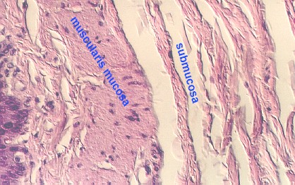 This figure shows a high magnification of the
muscularis mucosa and lamina propria (to the left of the muscularis mucosa). Also,
beneath the muscularis mucosa is a layer of connective tissue called the SUBMUCOSA.
This is seen to the right of the muscle layers in this photograph.
This figure shows a high magnification of the
muscularis mucosa and lamina propria (to the left of the muscularis mucosa). Also,
beneath the muscularis mucosa is a layer of connective tissue called the SUBMUCOSA.
This is seen to the right of the muscle layers in this photograph. 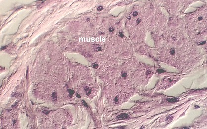 This figure shows a high magnification view of the
Muscularis mucosa.
This figure shows a high magnification view of the
Muscularis mucosa.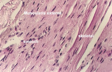

 This photograph shows a region containing the mucosa,
lamina propria, a bundle of the muscularis mucosa, and esophageal glands.
This photograph shows a region containing the mucosa,
lamina propria, a bundle of the muscularis mucosa, and esophageal glands.  This photo shows the abrupt transition from the
stratified squamous (left) of the esophagus to the simple cuboidal epithelium of the
stomach.
This photo shows the abrupt transition from the
stratified squamous (left) of the esophagus to the simple cuboidal epithelium of the
stomach.