 Graduate Microanatomy, 1998
Graduate Microanatomy, 1998
|
Digestive system study guide Lab Exercises:
Date page was last edited 06/06/04 |
Lab Exercises: Salivary Glands
1. Identify gland and duct cells in your slide and in these photographs.
Look at slide 28, the Submandibular Gland. This is a mixed salivary gland. Most of the glandular cells are serous. However some are mucous. 2. In the following photograph, identify mucous cells, serous cells and duct cells.
In the interlobar connective tissue, large EXCRETORY DUCTS can be seen. These are distinguished by their layers of nuclei and Goblet cells. The following photographs show views of this type of duct. Identify the Goblet cells.
3. Classify the epithelium in the EXCRETORY DUCTS____________________________
Slide 29 is the sublingual gland, which has more mucous cells than serous cells. This photograph shows a section of the sublingual gland. You can also see serous demilunes in this gland.
4. Consult your text and list the surface specializations found at the base of these striated duct cells. What is their function?
Higher magnification showing Goblet cells in the EXCRETORY DUCT of the Submandibular gland.
5. What is the difference between intercalated ducts and striated ducts?
Return to top of page | Course Design | Learning Aids | Learning modalities | |
 Look at slide 27, the Parotid gland. This is a
large salivary gland that consists of cells that are largely serous. It is divided
into lobes by connective tissue septa. Clusters of gland cells are organized in
Secretory acini. The cells are filled with granules. The saliva is collected in
small ducts that leave the lobules and move out into the connective tissue between the
lobes.
Look at slide 27, the Parotid gland. This is a
large salivary gland that consists of cells that are largely serous. It is divided
into lobes by connective tissue septa. Clusters of gland cells are organized in
Secretory acini. The cells are filled with granules. The saliva is collected in
small ducts that leave the lobules and move out into the connective tissue between the
lobes.  This view shows the granulated cytoplasm and the
smallest ducts leaving a cluster of glandular cells.
This view shows the granulated cytoplasm and the
smallest ducts leaving a cluster of glandular cells. 
 In some of the acini, the serous cells form a
cap (shaped like a half moon) over the centrally located mucous cells. This is
called a SEROUS DEMILUNE. These photographs show the granules in the serous glandular
cells as well as the mucous cells in the center of the demilune.
In some of the acini, the serous cells form a
cap (shaped like a half moon) over the centrally located mucous cells. This is
called a SEROUS DEMILUNE. These photographs show the granules in the serous glandular
cells as well as the mucous cells in the center of the demilune.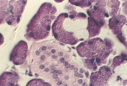


 This view shows one of the ducts in the sublingual
gland called the Striated duct. The nuclei have been pushed towards the lumen.
This view shows one of the ducts in the sublingual
gland called the Striated duct. The nuclei have been pushed towards the lumen. Slide 30 is a section of the submandibular gland
stained with Masson's trichrome and Alcian yellow. The yellow stain detects the
mucous cells. They are easy to distinguish.
Slide 30 is a section of the submandibular gland
stained with Masson's trichrome and Alcian yellow. The yellow stain detects the
mucous cells. They are easy to distinguish. This view shows a higher magnification of a serous
demilune in the submandibular gland.
This view shows a higher magnification of a serous
demilune in the submandibular gland.  Excretory ducts can also be found in slide 30.
They are distinguished by the the yellow staining Goblet cells.
Excretory ducts can also be found in slide 30.
They are distinguished by the the yellow staining Goblet cells. 
