


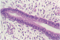


Below is a low magnification of a section through the ovary. The outer epithelium that covers the ovary. It is simple cuboidal and has inappropriately been named: GERMINAL EPITHELIUM. Also, note that as you move from the outer layers to the inner layers, there is an increasing complexity in the individual follicles. The outermost follicles, called primordial follicles appear to be a single oocyte in the center of a layer of simple squamous epithelium. The next layers have larger oocytes and more cuboidal epithelium. Subsequent layers have multiple layers of cuboidal epithelium. These represent various stages of follicular development which we will study in the following exercises. Note the increase in size of the oocyte.
Underneath the Germinal Epithelium is connective tissue called the "TUNICA ALBUGINEA" which consists of parallel arrays of collagen fibers. The next zone is called the CORTEX. It is distinguished by the ovarian follicles. The innermost zone which consists of loose connective tissue, is called the MEDULLA. Major blood vessels penetrate in this area in a region called the hilus. Why is the name "Germinal Epithelium" inappropriate?

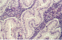
The above photograph shows another photo of the ovarian cortex. The outer layers of follicles are good examples of primordial follicles. The lower row of follicles are primary follicles with the large central oocyte and 1-more layers of cuboidal epithelium.
Primordial follicles
Find the germinal epithelium and primordial follicles which are in the the layer beneath The The simple squamous epithelium is the major diagnostic feature of these follicles seen below.


The following photograph is of a section through the cortex of a fetal ovary. Each female is born with all of the oocytes she will ever have in her lifetime.
Classify the follicles in the above section through the fetal ovary. The oogonia is arrested in a meiotic prophase.

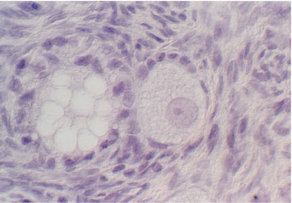
Primary follicle
When the epithelium around the oocyte becomes cuboidal, the follicle is called a "primary follicle".The following photograph shows a very early example (follicle to the right). The cuboidal cells are called "GRANULOSA CELLS" and they serve to help nourish and protect the oocyte. You can also see clusters of thin, fusiform cells condensing outside the follicles. These are the beginnings of the THECA INTERNA. Compare this view with Figures your text. IA ZONA PELLUCIDA is seen around the oocyte, which is a layer of glycoproteins through which the oocyte and granulosa cells communicate.
Atretic Follicle
Young Females begin their reproductive life with 400,000 primordial follicles. Fewer than 500 of these will complete maturation and be released. Most oocytes never advance to maturity. Even as early as during fetal development, the oocytes begin to degenerate and the process continues throughout adult life. These are called ATRETIC FOLLICLES. They may degenerate at any stage and your text describes different levels of degeneration, depending on the maturity of the follicle. Sometimes it is difficult to distinguish an atretic follicle from one in which the oocyte is poorly fixed. The best way to distinguish one is to look for a 'glassy membrane" appearance. Sometimes only a collapsed zona pellucida remains. The figure below is an illustration.

Secondary Follicle
As the follicle grows, the granulosa cells continue to proliferate until they are lined up in about 6-10 rows around the oocyte. Then, they develop a clear fluid region, called the ANTRUM. This is filled with substances that may protect, nourish, and stimulate the oocyte development. With the appearance of the antrum, the follicle becomes a SECONDARY FOLLICLE. The following photograph illustrates a secondary follicle. Compare it with the illustrations in your text. Identify the following structures in this photograph: primordial follicle, primary follicle, antral, or secondary follicle, zona pellucida, antrum, granulosa cells, theca interna cells.

As the follicle continues to grow, so does the antrum. Eventually, the oocyte is surrounded by a "hill" of granulosa cells called the CUMULUS OOPHORUS that projects into the antrum. This is shown by the following photographs.


Once you have identified the stages leading up to the largest, secondary follicles, try to find examples in other slides including slide 75, 1, and 78.. Each of these may have examples, although the fixation has not preserved the follicles well.
Graffian Follicle

The mature follicle is also called the GRAFFIAN FOLLICLE. These are not shown on the slides, because they are huge. Read the section in your text to gain an appreciation of the further growth and development. How large is the oocyte in the mature follicle? What stages of meiotic division are completed by the mature oocyte before ovulation? Is the product of that division, two mature oocytes? What hormone from the pituitary stimulates the development of the follicle?
Click here to study the post-ovulation events in the ovary.
Gwen V. Childs, Ph.D., FAAA
Department of Neurobiology and Developmental Sciences
University of Arkansas for Medical Sciences
4301 W. Markham, Slot 510, Little Rock, AR 72205
For questions or concerns, send email to this address
