


The fertilized ovum divides as it moves down the oviduct. By the fourth day, a central cavity appears and it is called the blastocyst. This attaches to the uterine endometrium which is in the secretory phase. After the blastocyst attaches, the endometrium undergoes the "Decidual reaction" The stellate stromal cells enlarge, and become polyhedral. They contain glycogen and lipid and presumably provide a source of nourishment for the implanting embryo.
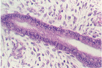
Slide 90 shows uterine epithelium undergoing the decidual reaction. The area is shown in the photos below. Pieces of placenta might be seen attached to the outer edges.


At one pole of the blastocyst, the inner cell mass develops. This will be the embryo. The rest of the blastocyst will form the placenta. This is called the TROPHOBLAST. After division, the trophoblast cells give rise to the CYTOTROPHOBLAST and a thick outer layer called the SYNCYTIAL trophoblast. The syncytiotrophoblast, as its name suggests, is a syncytium. It invades the endometrium and it eventually becomes completely encapsulated in the the endometrium Eroded maternal blood vessels provide nourishment and oxygen for the developing embryo and placenta.
What is the origin of the nuclei in the syncyiotrophoblast? What hormone is secreted by the placenta (and diagnostic of pregnancy)? It functions like which pituitary horrmone? (and is in fact chemically very similar).
At about day 15, the trophoblast shell sends villi deeper into the endometrium. These villi are filled with mesenchyme (called secondary villi) and will, by the end of the 3rd week, contain blood vessels (tertiary villi.) Maternal blood percolates slowly in the space created outside the villus. Look at slide 89 in your class slide set. The right hand section shows a young placenta. Look at the section with the low power. In the lower left of the section, there is a twisted, folded and otherwise distorted portion of the chorionic plate. It is amass of connective tissue covered by two types of epithelia. The side lined by flat epithelium is continuous with the amniotic sac. It faces the embryo. The side lined by two layers of trophoblast cells faces the uterus and gives rise to the villi. The photos below show the chorionic plate and related structures. The first photo shows the side facing the uterus.

The following photos show the portion facing the embryo. It is distinguished by the simple squamous epithelium. In the lower right corner, one can see the denser epithelium of the side facing the uterus.


Look at the side of the chorionic plate facing the uterus and look for stem villi originating from the chorionic plate. They are filled with mesenchyme and lined by the two layers of trophoblast cells. The inner layer, the cytotrophoblast, divides and gives rise to the outer layer, the syncytiotrophoblast. This is a multinucleated cellular layer that covers the surface of the villi.


Inside the villi, one can see blood vessels. However, note the distance between the vessel and the surface. This is the distance that nutrients and gases must cross to pass from the maternal blood (outside the villi) to the inside of the fetal blood vessels. The syncytiotrophoblast provides a cellular gateway through which all substances may or may not pass. Then after several layers of connective tissue, the gases must pass through the endothelium of the capillaries.

Both the maternal and fetal components are seen in the larger section on slide 89. As a landmark, look for the amniotic side of the chorionic plate. This actually shows the amniotic sac. Then, move into the chorionic plate and identify the stem villi originating from this plate. The following figure shows the amnionic side.

Look at the epithelium of the villi. The cytotrophoblast has disappeared and there is now one layer of syncytiotrophoblast. In places, the cytoplasm has thinned and the nuclei have accumulated in groups and clusters. These are called SYNCYTIAL KNOTS. Some of the villi show acellular pink-staining material called FIBRINOID which covers the surface. The accumulation is significant because it reduces maternal-fetal exchange.


Also, in mature placental villi are many more capillaries that lie closer to the surface. The following photographs show examples.


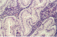
Look at the maternal side of the placenta, where ANCHORING VILLI can be seen embedded in uterine epithelia. The DECIDUAL BASALIS with the DECIDUA cells are found in the endometrium. Anchoring villi are shown in the following photographs.


The trophoblast has little smooth endoplasmic reticulum, yet it can synthesize both progesterone and estrogen. It works in partnership with what cells?
How does the placenta protect the fetus against invasion by T-lymphocytes?
Gwen V. Childs, Ph.D., FAAA
Department of Neurobiology and Developmental Sciences
University of Arkansas for Medical Sciences
4301 W. Markham, Slot 510, Little Rock, AR 72205
For questions or concerns, send email to this address
