
These photographs show parts of the wall of an elastic artery. It has been stained for elastin, so the elastic fibers show prominently. In the photograph to the left, the Tunica intima has a layer of endothelial cells as well as a subendothelial connective tissue layer. The next layer is the internal elastic layer which is usually counted as one of the dense elastic fiber lines seen in the photo (pick the densest, although it is counted more as a region, than a line). Note the elastic fibers in the Tunica media which also contains smooth muscle cells.
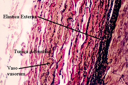
In the photograph to the right, the outer wall of the elastic artery is shown. The tunica media (not labeled) is seen separated from the Tunica adventia by the External elastic layer (Elastica Externa). Nerves and Vessels that supply the wall of the large vessel are seen in the tunica adventitia. Vessels are labeled (vaso vasorum).