Graduate
Microanatomy, 1998
Graduate
Microanatomy, 1998
Graduate
Microanatomy, 1998
|
Date page was last edited 06/06/04 |
Laboratory Exercises: Muscle Exercise 1. 1) Look at slide 11, which is a cross section of skeletal muscle. Describe (or diagram) the different levels of organization of the various components of skeletal muscle tissue. What type of tissue subdivides the components (name each layer)? 2) How does this organization contribute towards more efficient functioning of skeletal muscle? 3) Use the following figure to label the following components: perimysium, fasicle, muscle fiber
Exercise 2. 4) Slide 12 shows skeletal muscle in longitudinal section. Move your focus slightly up and down. What do you notice?_________________________________________ Is this a characteristic of skeletal muscle only__________________________? If not, where else do we find this characteristic_____________________? What contributes towards this appearance of skeletal muscle_____________________? 5) Where are the nuclei located_______________________________? An illustration of a longitudinal section of skeletal muscle is shown in the following figure.
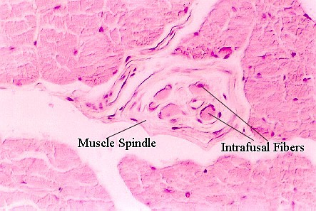
Slide 14 contains muscle spindles and the following photograph illustrates them.
10) What is the function of muscle spindles____________________________________________? Exercise 3. In your text, locate an electron micrograph which shows a longitudinal section of a skeletal muscle fiber. 6) Identify the structure(s) in skeletal muscle that send the signal from the plasma membrane (sarcolemma) to the sarcomere which ultimately stimulates contraction. ._______________________________________________ 7) Draw the basic "contractile unit" of skeletal muscle in a contracted and in an extended state. Exercise 4. 8) Look at the duodenum in cross-section. (Slide 53). Find the regions which contain muscle tissue. How can you distinguish muscle from connective tissue? ___________________________________________________________________________ 9) How is this muscle different from that seen in Exercises 1-3? _________________________________________________________________________ 10) How is this type of muscle organized?_________________________________________ 11) How does the organization contribute to the efficient transport of material through the GI tract?______________________________________________________________________ The following photograph illustrates smooth muscle in the GI tract.
Exercise 5. Look at slide 24, which shows a section through the skin of scalp. Find regions near hair follicles and identify bands of smooth muscle. These are "arrector pili" muscles. Be able to distinguish them from the surrounding connective tissue. The following photograph shows a view of Arrector pili muscles.
12. What is their function?__________________________________________________ Exercise 6. 13) Slide 15 shows cardiac muscle. Find an area with muscle fibers cut in longitudinal section. List features that distinguish cardiac muscle from skeletal muscle: ______________________________________________________________________________
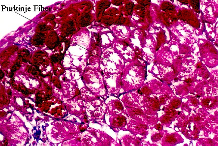
14) List the features that distinguish cardiac muscle from smooth muscle ________________________________________________________________________ 15) Identify the small dark lines running orthogonal to the direction of the fibers. What is the significance of these lines___________________________________________________________________? The following photograph shows longitudinally cut cardiac muscle fibers. 16) Slide 16 shows cardiac muscle that also contains Purkinje cells. Identify them and look at an electron micrograph of a Purkinje cell.
Why do the Purkinje cells look so empty?________________________________________ What is their function?________________________________________________________ Return to top of page | Course Design | Learning Aids | Learning modalities | |