Peripheral Nerve Histology

A section through peripheral nerve is shown. The differential staining shows the
different layers Blue stain indicates connective tissue. Outside
the nerve, loose connective tissue with adipose cells is called epineurium. The next layer
of connective tissue which is denser and ensheaths different bundles of nerve fibers is
called perineurium (shown dense blue in the slide). Delicate connective tissue surrounds
each nerve fiber; this is called endoneurium. In the photo and slide 3, you can see
endoneurium around each fiber as delicate blue fibers. A=Axon, M=myelin sheath. N=Node of Ranvier). Find the same structures on the following higher magnification of peripheral nerves.

The nerve fibers itself is stained purple/pink and is seen as a cylinder in the center of myelin which has a light orange stain. Use the above photograph to find the following:
- Node of Ranvier
- axon
- myelin sheath
- endoneurium.
The following is a section through a peripheral nerve stained with Osmium tetroxide to bring out the myelin.

The myelin sheath is stained because of the abundance of lipids in the membranes wrapping around the axon. Each myelinated axon looks like a donut. You can also see adipose cells well in this section. They contain stained, preserved fat droplets that stain dark brown-black.
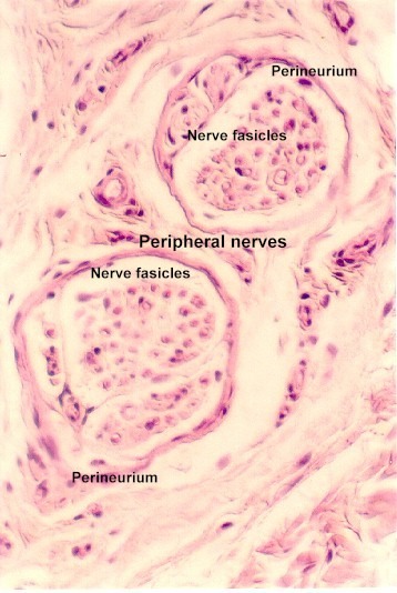
A peripheral nerve stained with Verhoeff's van Giesson stain. It shows nerve bundles called fasicles, epineurium, perineurium, endoneurium, axons and myelin. Practice your skills: find each of these structures and label the following figure.

A section of skin is a good place to find examples of peripheral nerves in the connective tissue. Learn to recognize these in connective tissue.

URL Address: http://microanatomy.net/nerve/peripheral_nerve_histology.htm
Gwen V. Childs, Ph.D., FAAA
Department of Neurobiology and Developmental Sciences
University of Arkansas for Medical Sciences
4301 W. Markham, Slot 510
Little Rock, AR 72205
For questions or concerns, send email to this address


