 Graduate Microanatomy, 1998
Graduate Microanatomy, 1998
|
Respiratory system study guide Lab Exercises:
Date page was last edited 06/08/04 |
Laboratory Exercises:
Respiratory system
Look at slide 44 This is a frontal section through one-half of the larynx. Follow the epithelial lining and note that it changes from Respiratory epithelium (pseudostratified columnar) to stratified squamous non-keratinixed epithelium. The stratified squamous epithelium signals that you are in the True Vocal Cord. The photograph to the left illustrates the epithelium. Move deeper under this epithelium. You will first encounter dense connective tissue. This is the Vocal Ligament. Continue to follow this tissue to a mass of muscle cut in cross section. This is the Vocalis muscle. It is illustrated in the photograph, below.
Continue to study slide 44 and find the following structures. First, the region which shows a sinus-like invagionation lined with Respiratory epithelium is called the Laryngeal ventricle. Just above this ventricle is the so-called "false vocal cord" which contains many mixed glands (serous and mucous glands).
Trachea
2. Define the term "respiratory epithelium".
3. Where do you find respiratory epithelium?
4. Identify the serous and mucous cells in the glands on your section and also on this photograph (to the left). How did you tell them apart?
5. What is the function of these glands?
6. What type of cartilage is found in trachea?
Return to top of page | Course Design | Learning Aids | Learning modalities | |
 Larynx
 1. What type of muscle is found in the vocalis muscle?
 Look at slide 45 which is a cross section through the trachea.
The luman is lined with ciliated Pseudostratified columnar epithelium with Goblet
cells. The Epithelium is supported by a basement membrane which rests on a
slightly condensed layer of connective tissue. The photograph to the left is a semithin
section of trachea embedded in plastic.
Look at slide 45 which is a cross section through the trachea.
The luman is lined with ciliated Pseudostratified columnar epithelium with Goblet
cells. The Epithelium is supported by a basement membrane which rests on a
slightly condensed layer of connective tissue. The photograph to the left is a semithin
section of trachea embedded in plastic.  This section comes from your slide 45.
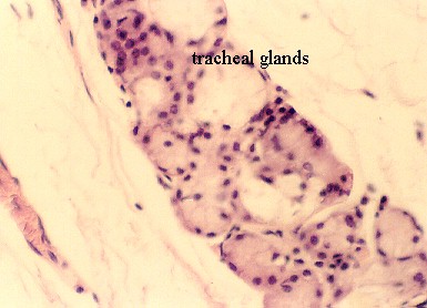 The connective tissue under the epithelium is called the
"adventitia". In this connective tissue are numerous seromucous (mixed)
glands, as well as blood vessels and nerves.
The connective tissue under the epithelium is called the
"adventitia". In this connective tissue are numerous seromucous (mixed)
glands, as well as blood vessels and nerves. 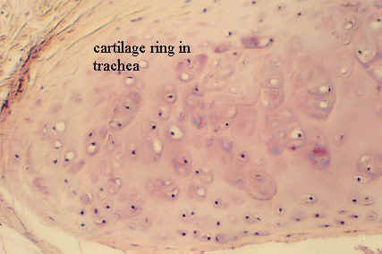 Note the C-shaped tracheal ring. You have already identified
perichondrium and the chondrocytes in this cartilage. You may find that the
cartilage is replaced by smooth muscle and elastic fibers in the
posterior wall of the trachea in your slide.
Note the C-shaped tracheal ring. You have already identified
perichondrium and the chondrocytes in this cartilage. You may find that the
cartilage is replaced by smooth muscle and elastic fibers in the
posterior wall of the trachea in your slide.