


Chondrocytes and Chondroblasts
Groups of 1-4 chondrocytes in hyaline cartilage. What are isogenous groups?
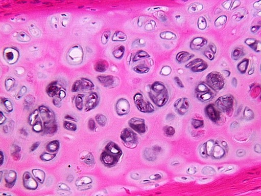
Study the perichondrium, outside cartilage, find layers of flattened cells that may give rise to chondrocytes as they begin to produce matrix. These are called chondroblasts. They are responsible for appositional growth.
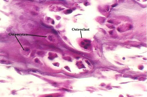
OSTEOBLASTS and OSTEOCYTES

The inner surface of bone is lined by osteoblasts, which are seen in the above photograph. This layer is called “endosteum”. Also evident are osteocytes in their lacunae and calcified cartilage.
Another bone cell important in remodeling is the Osteoclast, which are distinctive with multiple nuclei. They sits on the bone surface with multiple proceses, which increase surface area for absorption. Eventually, they form a depression called “Howship’s lacuna”. The next two photos show osteoclasts, osteoblasts and osteocytes.

URL Address: http://microanatomy.net/bone/cartilage_and_bone_cells.htm
Gwen V. Childs, Ph.D., FAAA
Department of Neurobiology and Developmental Sciences
University of Arkansas for Medical Sciences
4301 W. Markham, Slot 510
Little Rock, AR 72205
