
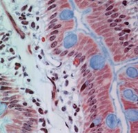

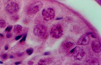


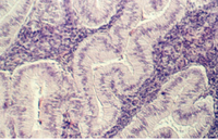

SIMPLE COLUMNAR EPITHELIUM
The epithelium is usually polarized and has a nucleus at the base of each cell. The following slide is from the intestine. The mucous is stained blue and large droplets are seen in one cell type (Goblet cell). Also, it is spread along the surface of the epithelium facing the lumen (inside of the intestine). What is its function? Mark the basal and apical boundaries of this epithelium.

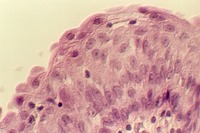
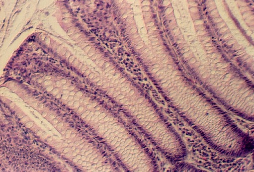
DUODENUM and JEJUNUM
In the photo from your duodenum slide an important apical surface specialization is
shown. This is called the brush border. What does it look like at the electron
microscopic level and what is its function? Also, can you see a droplet of mucous being
secreted? For more views of the intestinal epithelia see:
http://microanatomy.net/digestive/intestine.htm

The colon is shown below. Its columnar epithelium has many more Goblet cells. You can define the base of the cell by identifying the nuclei which are in a row beneath the mucous droplet.
URL Address: http://microanatomy.net/epithelia/simple_columnar.htm
Gwen V. Childs, Ph.D., FAAA
Department of Neurobiology and Developmental Sciences
University of Arkansas for Medical Sciences
4301 W. Markham, Slot 510
Little Rock, AR 72205
For questions or concerns, send email to this address
