
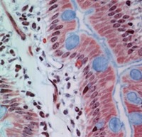


INTESTINE
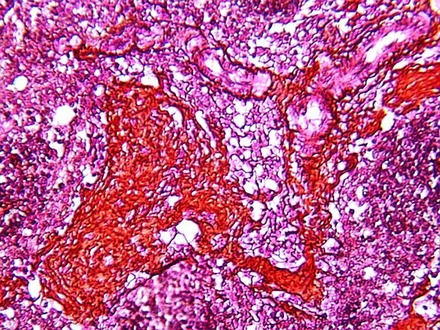
Loose connective tissue, also called Areolar is often used to link epithelia to other parts of
the organ wall. This is a section through the duodenum slide. Epithelia is shown on the
right and the remainder of the field is loose connective tissue (called "lamina
propria". Note that it is well vascularized and supplies the nutrients and
controlling factors for the surrounding cells, including epithelia. You can also see
strands of smooth muscle in the connective tissue. Can you identify a blood vessel (cut in
longitudinal section). It is a capillary...what type of epithelia lines this
capillary?
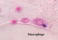
Note how many cells there are in the field of loose connective tissue, including the fibroblasts and fibrocytes that make the matrix and collagen fibers. Bundles of collagen fibers and spindle-shaped fibroblasts can be seen in the fields. Often the fibroblasts can be identified only by their long slender nuclei running parallel to the bundles of collagen fibers. They are seen as cells with the very thinnest nuclei, but they should not be confused with endothelial cells, or smooth muscle cells that have much fatter nuclei. See also photo of dense irregular and regular connective tissue.

Loose connective tissue is also found under the epidermis (epithelium) of the skin in a region called the dermis. You can distinguish it by its vascularity and cellularity (lots of cells and blood vessels). Look at your slide 25 and find the highly vascular area under the epidermis. The following photo shows thick skin.

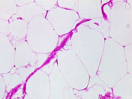
Adipose tissue is found in the hypodermis of the skin, or surrounding many organs muscles, nerves, etc. You can see it at the base of the section in slide 25. It is made of relatively large cells that are distinguished by a thin process surrounding a large droplet of fat. The nucleus is very thin and small. Thus, adipose tissue looks like a "honeycomb" with the cells being the walls of the chambers and the fat droplets filling the center. The processing of this section has removed the fat droplet and the cell center looks clear. If you want to see fixed, stained fat droplets, look at slide 8 and find the adipose tissue around the peripheral nerve.
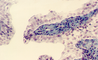
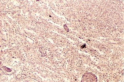
Loose connective tissue is also filled with reticular fibers which are fine fibers used for support. The following photos show regions from the spleen with reticular fiber stains. Note the fine bundles of fibers stained black. They help to provide the structural support for the cells and vessels in the loose connective tissue..

URL Address: http://microanatomy.net/connective_tissue/loose_connective_tissue.htm
Gwen V. Childs, Ph.D., FAAA
Department of Neurobiology and Developmental Sciences
University of Arkansas for Medical Sciences
4301 W. Markham, Slot 510
Little Rock, AR 72205
For questions or concerns, send email to this address
