

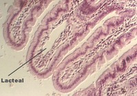

The intestine is specialized for absorption and further digestion, as well as water conservation. The mucosal epithelium continues to be simple columnar. However, base of the gland is called the "crypt" and there are no pits. Look at slide 53, which is the duodenum. Find the epithelium which is lining long projections called VILLI. The core of the villus contains the LAMINA PROPRIA and strands of smooth muscle, which is part of the MUSCULARIS MUCOSA. The following photo shows duodenal villi. Note the LACTEAL in the lamina propria. What type of vessel is the lacteal?

Mucosa
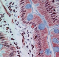
The epithelium of the duodenum consists of the COLUMNAR ABSORPTIVE CELLS interspersed with GOBLET CELLS. In the following photograph, these cells are evident along with their BRUSH BORDER. Underneath the epithelium, the lamina propria can be seen. Identify these cells and regions on the appropriate photographs:
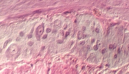
The following photo shows a better view of the lamina propria which is the core of the villi. Strands of smooth muscle, vessels, and numerous cells typical of connective tissue are found in this region.

Crypt of Lieberkuhn and Brunner's glands
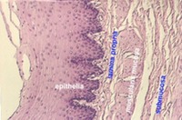
The base of the villus is called the CRYPT of LIEBERKUHN or INTESTINAL CRYPT. Under the crypts, you can find more lamina propria, strands of MUSCULARIS MUCOSA, and glands that distinguish the esophagus called BRUNNER'S GLANDS. These glands produce mucus and most of the glandular portion is located in the submucosa. Can you find a bundle of MUSCULARIS MUCOSA in the following photograph? The submucosa is the red connective tissue, seen best in the left of this photo.

Muscularis Externa and Auerbach's plexus
Identify the MUSCULARIS EXTERNA in slide 53. This consists of inner circular and outer longitudinal layers. Auerbach's plexus lies between the muscle layers, as shown in the following photographs.


Pancreatic duct and Common Bile duct
A portion of the pancreas is included on your slide 53. Two smaller hollow structures may be seen. These are the BILE DUCT and the PANCREATIC DUCT. The BILE DUCT wall contains circularly arranged smooth muscle and the lumen shows many irregular clefts and pockets. It is lined with a simple columnar epithelium. There is no muscularis mucosae. The photo below shows the Bile duct.

The pancreatic duct may not be as easy to find. It is lined with a simple columnar epithelium, but its lumen shows a much simpler outline and its wall contains only an inconspicuous amount of smooth muscle. Share with your neighbors if your section 53 does not contain a pancreatic duct.
What do the pancreatic and bile ducts carry to the duodenum lumen?
Jejunum and Ileum
After you find the above structures on slide 53, look at slides 54 and 55. The JEJUNUM, slide 54, shows the PLICAE CIRCULARES studded with VILLI. It is very important to identify the regions here so you can understand how much of the wall is involved in the major folds. The villi are lined by absorptive epithelium. What connective tissue is the core of the villi? What connective tissue is found in the Plicae circulares?
Paneth cells and Enteroendocrine cells
The muscularis mucosae is well developed and can be seen at the base of the villi.
It follows the folds of the Plicae circulares. Look at the base of the
glands, the Crypts. You can see clearly the granulated PANETH CELLS distinguished by
red granules pointed towards the lumen of the gland. You may also see
ENTEROENDOCRINE cells at the base of the glands. Examples of both types of cells are seen
in the following photo. Recall that the red granules of the enteroendocrine cells are
pointed towards the blood vessels in the lamina propria. 
Ileum
Look at slide 55, the Ileum. The MUCOSA extends into the lumen again in the form of long villi. Note that there are more GOBLET cells in the Ileum than in the Duodenum. You can also see clusters of LYMPHOID TISSUE, called PEYER's PATCHES. You might be able to find enteroendocrine cells, but the Paneth cells are not obvious. Also, find the SUBMUCOSA, MUSCULARIS EXTERNA and Auerbach's plexus.
Colon
The colon is characterized by mucosal folds that are no longer called villi. These are lined by many GOBLET CELLS and fewer ABSORPTIVE CELLS. The glands are shorter in the colon than in the small intestine. There are no Paneth cells.
Beneath the epithelium, you can see lamina propria. Other layers of the colon are similar to the submucosa and muscularis of the rest of the gut. However, the colon has muscle layers arranged in TAENIAE COLIA.


The above photograph shows a higher magnification of the colon epithelium with its numerous Goblet cells. Compare this view with that showing the SURFACE MUCOUS CELLS of the stomach.
Appendix
The epithelium of the Appendix contains numerous Goblet cells, but they are not as numerous ad in the colon or rectum. There are no Villi or Plicae. The lamina propria and submucosa is filled with diffuse lymphoid tissue. Note that, unlike Peyer's patches in the ileum, the follicles in the appendix completely surround the lumen. Also, unlike the ileum, there is debris in the lumen.


You may find a number of ENTEROENDOCRINE CELLS in the appendix. As shown in this photograph, the granules are pointed in the direction of the blood vessels in the lamina propria. You can see eosinophils in the lamina propria as well.
Gwen V. Childs, Ph.D., FAAA
Department of Neurobiology and Developmental Sciences
University of Arkansas for Medical Sciences
4301 W. Markham, Slot 510, Little Rock, AR 72205
For questions or concerns, send email to this address
http://microanatomy.net/digestive/intestine.htm
